
I remember the moment clearly. It was a Thursday afternoon last month when Rebecca walked through our doors at 50 Lacoste Blvd, carrying her twelve-year-old cat, Oliver. He hadn’t been himself for two days—refusing food, hiding under the bed, and crying when she tried to pick him up. The worry in her eyes was the same look I’ve seen hundreds of times over my years in veterinary medicine. She needed answers, and she needed them quickly.
Twenty minutes later, we had those answers. Digital radiography revealed a bladder stone the size of a marble, and an ultrasound confirmed there was no kidney damage. Oliver went home that same afternoon with a treatment plan, scheduled surgery for the following week, and Rebecca left with something precious in veterinary care—certainty instead of anxious waiting.
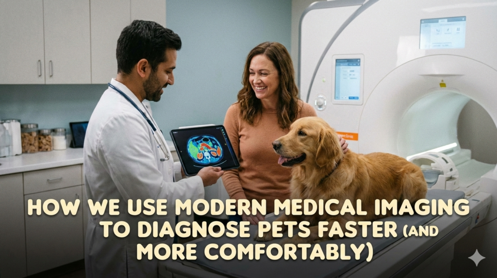
This is what modern medical imaging has brought to veterinary medicine, and it’s changed everything about how we diagnose and treat our patients at Lacoste Animal Hospital.
The Evolution From Film to Digital: Why It Matters to Your Pet
Ten years ago, taking radiographs meant waiting. We’d position the patient, expose the film, develop it in chemical baths, wait for it to dry, and then finally examine the images. The entire process could take thirty to forty-five minutes, and that’s if everything went perfectly on the first attempt. If we needed a different angle or if the exposure wasn’t quite right, we’d start over.
Today, when you bring your pet to our facility for imaging, the experience is completely different. Our digital radiology system captures images instantly. Within seconds of taking the radiograph, it appears on high-resolution monitors where we can zoom in, adjust contrast, and examine every detail. What used to require multiple exposures and patient repositioning now often happens with a single, quick capture.
This speed isn’t just about convenience. For anxious animals, every minute on the table matters. The faster we work, the less stressful the experience becomes. When you’re searching for “radiology near me” or “medical imaging near me,” understanding this difference between traditional and modern facilities should factor into your decision.
Inside Our Medical Imaging Suite
The medical imaging area at our hospital represents a significant investment in technology and training. Walk into this space, and you’ll notice it feels different from a typical exam room. The lighting is controlled, the equipment gleams with the hallmarks of modern veterinary care, and most importantly, the atmosphere remains calm and quiet—designed specifically to keep pets comfortable during procedures.
Our in-house medical imaging capabilities center around two primary technologies: digital radiography and diagnostic ultrasound. Together, these tools allow us to see inside your pet’s body without invasive procedures, providing critical information that guides treatment decisions.
The digital X-ray system mounts on a table that can be adjusted for patients of all sizes, from tiny kittens to large breed dogs. The ultrasound machine sits on a mobile cart, allowing us to bring it directly to patients in exam rooms when needed, particularly useful for animals too sick or anxious to move to the imaging suite.
Digital Radiography: Seeing Bones, Organs, and Everything Between
When most people think about X-rays, they imagine looking at bones. And yes, radiography excels at showing us skeletal structures. Fractures, arthritis, bone tumors, and developmental abnormalities all appear clearly on radiographs. But modern digital imaging reveals so much more.
Last Tuesday, a worried owner brought in Duke, a seven-year-old German Shepherd who had suddenly stopped eating and seemed uncomfortable. Physical examination suggested something was wrong in his abdomen, but we needed more information. Radiographs revealed the problem immediately—a corn cob lodged in his intestines, causing a complete blockage.
Without medical imaging, we would have been guessing. With it, we knew exactly what we were dealing with, where the problem was located, and how urgently we needed to intervene. Duke went straight to surgery, and the corn cob was removed before any permanent damage occurred. Three days later, he was back to his enthusiastic, food-loving self.
This is the power of having access to quality medical imaging right here in Brampton. When pet owners don’t have to drive to a distant facility or wait days for an appointment at a specialized ultrasound clinic, diagnosis happens faster and treatment begins sooner.
The Ultrasound Advantage: Real-Time Internal Viewing
While radiography gives us static images of dense structures, ultrasound provides something entirely different—real-time video of soft tissues and organs in motion. Watching a heart beat on the ultrasound screen, seeing blood flow through vessels, observing kidney structure, or identifying fluid accumulations gives us information that radiographs simply cannot provide.
The technology works using high-frequency sound waves that bounce off tissues and return to create images. It’s completely non-invasive, doesn’t require sedation in most cases, and carries zero radiation exposure. For pregnant animals, anxious patients, or pets with conditions where sedation poses risks, ultrasound often becomes our first choice for internal imaging.
Take the case of Maggie, a middle-aged Beagle who came in because her owner noticed her drinking excessive amounts of water. Blood work from our in-house diagnostics showed elevated kidney values, but we needed more information. An abdominal ultrasound revealed one kidney was significantly smaller than the other, with changes consistent with chronic kidney disease. This finding completely changed her treatment approach and prognosis discussions with her owner.
The beauty of having an ultrasound machine on-site means we can perform these examinations during regular appointments. No need to schedule a separate visit to a specialty imaging ultrasound facility. No waiting weeks for an opening. We simply walk to the ultrasound machine and get answers.
Comparing Old and New: What Pet Owners Gain
The difference between traditional and modern veterinary medical imaging extends beyond just speed. Image quality has improved dramatically. Digital sensors are more sensitive than film ever was, meaning we can use less radiation while still capturing excellent images. The ability to manipulate digital images—adjusting brightness, contrast, and magnification—means we can extract more information from each radiograph.
Consider how this plays out in practice. When we take a chest radiograph looking for heart disease or lung problems, we can adjust the image to emphasize either the soft tissues of the lungs or the bony structures of the ribs and spine without taking additional exposures. With film, we’d need to take multiple radiographs with different settings, exposing the patient to more radiation and extending the examination time.
Medical imaging consultants in human medicine have long recognized these advantages. Veterinary medicine has now caught up, and facilities like ours bring hospital-grade imaging to companion animal care. When you’re looking for quality care and searching for a “vet near me,” asking about imaging capabilities should be part of your evaluation process.
The Comfort Factor: Minimizing Stress During Imaging
I’ve seen countless pets over the years, and I can tell you with certainty that they pick up on anxiety. When imaging procedures dragged on for forty-five minutes with multiple repositionings and adjustments, animals became increasingly stressed. That stress sometimes required sedation, which added risk and recovery time to what should have been a simple diagnostic procedure.
Modern imaging has changed this dynamic completely. Most radiographs now take less than five minutes from start to finish. We position the patient, capture the image, verify it’s diagnostic quality, and we’re done. Many pets barely realize what happened before they’re getting treats and heading back to their owners.
For ultrasound examinations, we’ve refined our approach over years of practice. Most dogs and cats tolerate abdominal ultrasound remarkably well. We place them on a comfortable padded table, apply warm gel to their belly, and gently move the probe across their skin. The vast majority of patients relax into the process, especially when a veterinary technician is providing gentle reassurance and the occasional treat.
There are exceptions, of course. Some patients need sedation for quality imaging, particularly if we’re examining painful areas or if the pet is too anxious to hold still. But those cases have become the exception rather than the rule. The speed and ease of modern medical imaging means most pets handle diagnostic procedures far better than they did a decade ago.
Exotic Pets and Special Imaging Considerations
When people search for an “exotic vet near me,” they’re often worried about finding someone with appropriate experience and equipment for their unusual companion. Imaging exotic pets presents unique challenges. A rabbit’s skeleton is entirely different from a dog’s. A bird’s respiratory system doesn’t look anything like a mammal’s. Reptiles have completely different anatomy.
Our team has experience with all of these cases. Last month, we examined a pet rabbit who had stopped eating. Radiographs revealed dental disease—a common problem in rabbits that can’t always be seen during a physical exam. Two weeks ago, a concerned owner brought in a bearded dragon who was lethargic and not moving well. Radiographs showed metabolic bone disease, a condition caused by nutritional imbalances that weakens the skeleton.
Having imaging capabilities in-house means exotic pet owners don’t have to drive across the region searching for facilities willing and able to image their unusual companions. We have the equipment, the experience, and the willingness to work with all types of pets at our location on Lacoste Blvd.
When Imaging Leads to Life-Saving Interventions
Some imaging studies provide interesting information that guides long-term management. Others reveal problems requiring immediate intervention. These emergency situations showcase why having advanced medical imaging available during extended hours matters so much.
It was a Friday evening last fall, around 8 PM, when Tom rushed through our doors carrying Bailey, his young Labrador who had been fine that morning but suddenly collapsed while playing fetch. Bailey was weak, her gums were pale, and her heart rate was racing. Abdominal ultrasound revealed free fluid in her abdomen—she was bleeding internally.
Within minutes, we had identified a ruptured splenic mass as the source of bleeding. Bailey went immediately to surgery. Without rapid ultrasound imaging, we would have been operating blind, unsure of what we’d find or even where to look first. The imaging gave us a roadmap, and that made all the difference. Bailey recovered beautifully and is still chasing tennis balls two years later.
These are the moments when medical imaging transcends being merely useful and becomes absolutely critical. When searching for veterinary care, especially if you have a breed prone to certain conditions, knowing a facility has comprehensive imaging immediately available can literally save your pet’s life.
The Integration of Imaging with Complete Diagnostic Care
Medical imaging doesn’t exist in isolation. It works best when integrated with other diagnostic tools. At Lacoste Animal Hospital, we combine imaging with comprehensive blood work, urinalysis, and other laboratory tests to build complete diagnostic pictures.
Consider the diabetic cat who comes in for monitoring. Blood work tells us about glucose control and kidney function. Radiographs might reveal bladder stones, a common complication in diabetic cats. Ultrasound can show us whether the kidneys show any changes from chronic high blood sugar. Each piece of information adds to our understanding, and modern imaging provides crucial pieces of that puzzle.
This integrated approach to diagnostics is what sets comprehensive veterinary facilities apart from basic clinics. When you need insight into your pet’s health, having access to multiple diagnostic modalities under one roof means faster answers and more coordinated care.
Real Patient Stories: How Imaging Changed Outcomes
Beyond the dramatic emergency saves, medical imaging impacts everyday cases in profound ways. Let me share a few examples that illustrate this point.
Sophie, an eight-year-old mixed breed dog, came in for routine wellness and preventive care. Her owner mentioned she’d been coughing occasionally but thought it was just excitement. A precautionary chest radiograph revealed early heart enlargement. We caught the condition before it progressed to heart failure, started medication, and Sophie continues to thrive three years later.
Then there’s Charlie, a cat who was losing weight despite eating well. Abdominal ultrasound revealed intestinal lymphoma. While the diagnosis wasn’t what we hoped for, having definitive answers allowed his owners to make informed decisions about treatment. Charlie responded well to chemotherapy and enjoyed an additional eighteen months of quality life.
These stories represent the daily reality of modern veterinary imaging. It’s not always about dramatic rescues. Often, it’s about finding answers, providing clarity, and enabling better treatment decisions.
Understanding Image Interpretation: The Human Element
All this technology means nothing without trained professionals who know how to interpret what they’re seeing. Reading radiographs and ultrasound images requires years of training and experience. Subtle changes in organ size, unusual fluid patterns, or early signs of disease can be easy to miss if you don’t know what to look for.
Our veterinarians have undergone extensive training in diagnostic imaging. Some have completed additional continuing education programs focusing specifically on radiology and ultrasound. We also maintain relationships with veterinary radiologists who can review complex cases and provide specialist interpretation when needed.
This combination of in-house expertise and access to specialized medical imaging consultants means we can handle everything from routine radiographs to complex cases requiring expert review. Pet owners get the benefit of both immediate on-site interpretation and specialist consultation when cases warrant it.
Frequently Asked Questions About Pet Imaging
-
Does my pet need sedation for radiographs or ultrasound?
Most pets don’t require sedation for standard imaging. Digital radiography is so quick that most animals tolerate positioning for the few seconds needed to capture images. Ultrasound is generally well-tolerated, though some pets need mild sedation if they’re painful, anxious, or we need them to remain very still for detailed cardiac imaging.
-
Is radiation from X-rays dangerous for my pet?
Modern digital radiography uses significantly less radiation than older film-based systems. The exposure from a typical radiograph series is minimal, and the diagnostic benefit far outweighs any theoretical risk. We follow the principle of using the minimum necessary exposure to obtain diagnostic images.
-
How long do imaging procedures take?
Simple radiograph studies typically take five to ten minutes. More complex examinations might require fifteen to twenty minutes. Ultrasound examinations vary depending on what we’re examining, but most take fifteen to thirty minutes. Unlike some facilities that schedule imaging days or require separate appointments, we often complete imaging during regular visits.
-
Will I get copies of my pet’s images?
Yes, we can provide digital copies of radiographs and ultrasound images if requested. Many pet owners appreciate having these records, especially if seeking second opinions or transferring to new veterinarians.
-
What conditions can you detect with imaging?
The list is extensive—fractures, arthritis, bladder stones, foreign objects, organ enlargement or shrinkage, tumors, pregnancy, heart disease, lung conditions, fluid accumulations, intestinal blockages, and many other conditions. Imaging provides invaluable information about internal health that physical examination alone cannot reveal.
-
Do you refer cases to specialist imaging facilities?
For most cases, our in-house capabilities are sufficient. Occasionally, we recommend advanced imaging like CT scans or MRI, which require referral to specialty facilities. However, these cases are relatively uncommon. The vast majority of pet imaging needs can be met right here.
Access and Availability: When You Need Imaging
Location and availability matter when you need diagnostic imaging. Having to drive an hour to a specialty ultrasound clinic, then wait days or weeks for an appointment, creates unnecessary delays in diagnosis and treatment. This is particularly problematic for urgent cases where timing impacts outcomes.
Our location at 117, 50 Lacoste Blvd in Brampton makes us accessible to pet owners throughout the region. Our extended hours—open until 9 PM on Mondays, until 10 PM on Fridays, Saturdays, and Sundays—mean imaging is available when you need it, not just during traditional business hours.
If you notice concerning symptoms in your pet and need medical imaging, you can reach us at (905) 913-8888 to discuss whether your pet needs to be seen urgently or if a scheduled appointment makes sense. For non-urgent matters, emailing petcare@lacosteanimalhospital.ca allows our team to respond with scheduling options.
The Future of Veterinary Medical Imaging
Technology continues advancing, and veterinary imaging evolves alongside human medical imaging. Three-dimensional reconstruction, advanced Doppler ultrasound for detailed blood flow assessment, and artificial intelligence assistance in image interpretation are all on the horizon or already emerging in veterinary medicine.
We stay current with these advances through continuing education, equipment updates, and careful evaluation of new technologies. The goal remains providing Brampton pet owners with the best diagnostic capabilities available while maintaining the personal, caring approach that defines our practice.
Why Imaging Capability Should Guide Your Veterinary Choice
When choosing where to take your pet for care, many factors come into play—location, hours, staff personality, and cost all matter. But diagnostic capability should rank high on that list. A veterinarian without adequate imaging tools is working with one hand tied behind their back, forced to make educated guesses rather than informed decisions.
This doesn’t mean every clinic needs every piece of high-tech equipment. But in 2026, digital radiography and ultrasound should be considered standard tools, not luxury additions. When searching for a “vet near me” or researching facilities, asking about imaging capabilities provides insight into how comprehensively a clinic can investigate health concerns.
Making Imaging Work for Your Pet
Understanding what modern medical imaging can do helps pet owners make informed decisions about their animal’s care. When your veterinarian recommends radiographs or ultrasound, that recommendation comes from knowing these tools provide information essential for proper diagnosis and treatment planning.
Some pet owners initially hesitate at imaging recommendations, concerned about cost or necessity. It’s a fair question to ask what information imaging will provide and how it will change treatment approaches. Good veterinarians should be able to explain clearly why they’re recommending specific diagnostic tests and what they hope to learn.
In our experience, most pet owners appreciate having clear answers rather than treating blindly and hoping for the best. Medical imaging provides those answers, often revealing conditions that would have progressed undetected until they became far more serious and difficult to treat.
Bringing It All Together
Modern medical imaging has transformed veterinary medicine in ways that benefit every pet owner. Faster diagnoses mean quicker treatment starts. Better image quality means more accurate diagnoses. Less stressful procedures mean happier pets. And having comprehensive imaging available in-house means no delays, no driving across the region, and no juggling multiple appointments at different facilities.
This is what we offer at Lacoste Animal Hospital—medical imaging that meets the standards you’d expect from a human hospital, delivered with the compassion and personalized attention your pet deserves. Whether you’re dealing with an emergency on a Saturday night or bringing your senior pet in for routine screening, our imaging capabilities stand ready to provide the answers you need.
The veterinary landscape has changed dramatically over the past decade. Facilities once considered cutting-edge for having basic X-ray capability now seem outdated compared to what’s possible with modern digital imaging and ultrasound. We’re proud to bring these capabilities to Brampton, making advanced diagnostic tools accessible to local pet owners without requiring referrals to distant specialty centers.
When your pet needs answers, when symptoms don’t make sense, when treatment isn’t working as expected, or when you simply want thorough screening for peace of mind, medical imaging provides clarity. And in veterinary medicine, clarity leads to better outcomes, fewer complications, and pets who live longer, healthier lives.
That’s why we’ve invested so heavily in imaging technology and training. That’s why we maintain equipment to hospital-grade standards. And that’s why we make these capabilities available during extended hours throughout the week. Your pet’s health deserves nothing less than the best diagnostic tools modern veterinary medicine can provide.
The next time you’re concerned about your pet’s health, remember that getting answers doesn’t have to mean days of waiting or driving to multiple facilities. Quality medical imaging is available right here in your community, delivered by professionals who combine technical expertise with genuine care for your animal’s wellbeing. That combination—advanced technology in caring hands—represents the future of veterinary medicine, and that future is available today at our facility on Lacoste Blvd.